IB Biology Sub-topic B2.3 Notes
Surface-to-Area Volume
Now that you know about cell specialization, you are expected to understand the several adaptations cells develop to better perform their function. This begins with adaptations for an increased surface-to-volume ratio. This is typically done by:
- Reducing cell diameter
- Flattening or elongating the cell
- Making large membrane invaginations
- Forming hair-like structures called microvilli
Using this, cells shaped like or cylinder with microvilli or invaginations will have a greater surface area than a sphere of the corresponding volume. Two examples you need to remember are:
- The microvilli contained in luminal cells of the proximal convoluted tubule. These maximize surface area for efficient diffusion of substances whilst maintaining cell shape to form a tube.
- The flattened disc shape of erythrocytes with a large invagination on either side. This increases surface area for oxygen to diffuse and bind to haemoglobin.
Alveoli
Next, alveoli. They contain several important adaptations to effectively perform gas exchange:
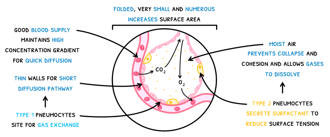
- Alveoli are folded, very small and very numerous, increasing the surface area for gas exchange.
- They have a good blood supply, maintaining a high concentration gradient to allow gasses to diffuse more quickly.
- They contain Type I pneumocytes, which have extremely thin walls to provide a short diffusion pathway. They thus act as the site of gas exchange.
- They contain Type II pneumocytes, which which secrete a solution known as surfactant. This makes the alveoli moist to:
- Reduce surface tension.
- Prevent collapse and cohesion of the walls.
- Allow gasses to dissolve and passively diffuse into the blood.
Muscle cells
Next, muscle cells. Humans contain three types of muscle: smooth muscle found in the alimentary canal, cardiac muscle found in the heart, and skeletal muscle found in classic muscle.
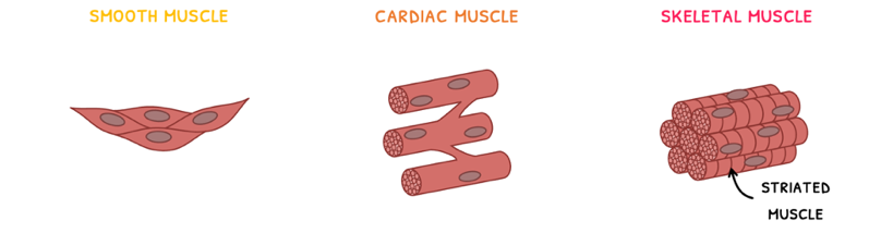
Skeletal muscle, also known as striated muscle, is easily recognizable due to its striations. These striations are a result of its structure. The tissue is composed of many muscle fibers bundled together into fasciculi. Each muscle fiber is formed from the fusion of embryonic muscle cells – hence why each muscle fiber is multi-nucleated. You are expected to know the detailed structure of a muscle fiber. It is composed of:
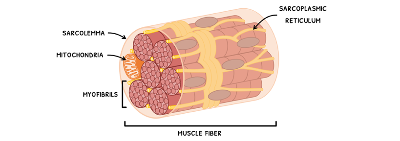
- A sarcolemma – the plasma membrane of the muscle fiber.
- Myofibrils – these are the long contractile fiber organelles of a muscle fiber.
- Sarcoplasmic reticulum – this is a network of tubules within the muscle fiber that stores calcium ions and acts to coordinate contraction across the whole fiber.
- Mitochondria – in abundant supply, these provide the large amounts of ATP necessary for contraction.